#How to read mri images of right knee full#
However, most medical professionals recommend a follow-up appointment to discuss the medical jargon contained in radiology reports for a full understanding of them. In addition to safeguarding patient information and improving efficiency, the advent of electronic health records (EHRs) – also called electronic medical records (EMRs) – has given patients access to their radiology reports electronically through secure online portals. One study found that a delay in delivering the results of a scan can cause patients to experience anxiety, as they assume the delay indicates that something bad was found in the scans. Releasing imaging results too early – when not enough examination and analysis has been done – or too late can have negative effects on patient experience.

The mode of transmission between practice to doctor (e.g., fax).Whether a previous X-ray or another image is being compared to it.Whether more information is required before the radiologist can properly interpret the images.The urgency with which the results are needed.The speed at which you receive the results of an X-ray will depend on: There are many factors that come into play regarding turnaround time. What Can Make My Scanning Results Faster? Patients who are having imaging tests due to accidents or surgeries are usually the first ones served – this is called the triage process (pronounced TREE-ahj). Oftentimes, large academic hospitals have a high volume of results to analyze and process. The results are “read” by a radiologist, and the turnaround time for their analysis depends on their availability and where the imaging study was done. The results from an MRI scan are typically interpreted within 24 hours, and the scans themselves are usually given immediately to the patient on a disc after the MRI is complete. The swift transmission of diagnostic information is important to both patients and referring physicians.


How the Medical Industry Is Trying to Speed Up Imaging ResultsĪccording to the Journal of the American College of Radiology, imaging providers across the United States are searching for ways to shorten the turnaround time for compiling and delivering results. The doctor or specialist will then relay the full analysis to you, along with recommendations and/or prescriptions. The radiology report – the written analysis by the radiologist interpreting your imaging study – is transmitted to the requesting physician or medical specialist. If you’ve ever had a diagnostic imaging study done – such as an X-ray, MRI, ultrasound, or CT scan – you may understand what it feels like waiting for those results. So, it’s no surprise that when it comes to medical imaging, fast turnaround time is expected by doctors and their patients. There is no significant joint effusion.In today’s day and age, you can find a walk-in urgent care clinic in virtually every neighborhood, and you can even get your flu shot at the nearby pharmacy. The quadriceps and patellar tendons are intact. There are mild degernative changes int he lateral compartment, with partial thickness articular surface cartilage thinning and adjacent marginal osteophyte formation. Lateral Meniscus: There is a complex tear along the anterior horn, body and posterior horn of the lateral meniscus. Medial Meniscus: The medial meniscus is intact. LCL: The lateral collateral ligament is normal. MCL: The medial collateral ligament is intact.

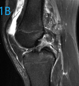
PCL: The posterior cruciate ligament is intact. Technique: Multi-planar images of the right knee were obtained at 1.5 Tesla without IV contrast.ĪCL: The anterior cruciate ligament is intact. The MRI Knee Case Study procedure included multi-planar images of the right knee obtained without IV contrast on our 1.5 Tesla MRI machine. Our case study of the month is an MRI scan of the right knee with history of internal derangement.


 0 kommentar(er)
